The Florey Institute
  • Home
  • News and Events
  • Research
  • The Team
  • Antimicrobial Resistance MSc
  • Contact

Day 10 - Seeing DNA and Bacteria without 'sight'

17/3/2019

2 Comments

 
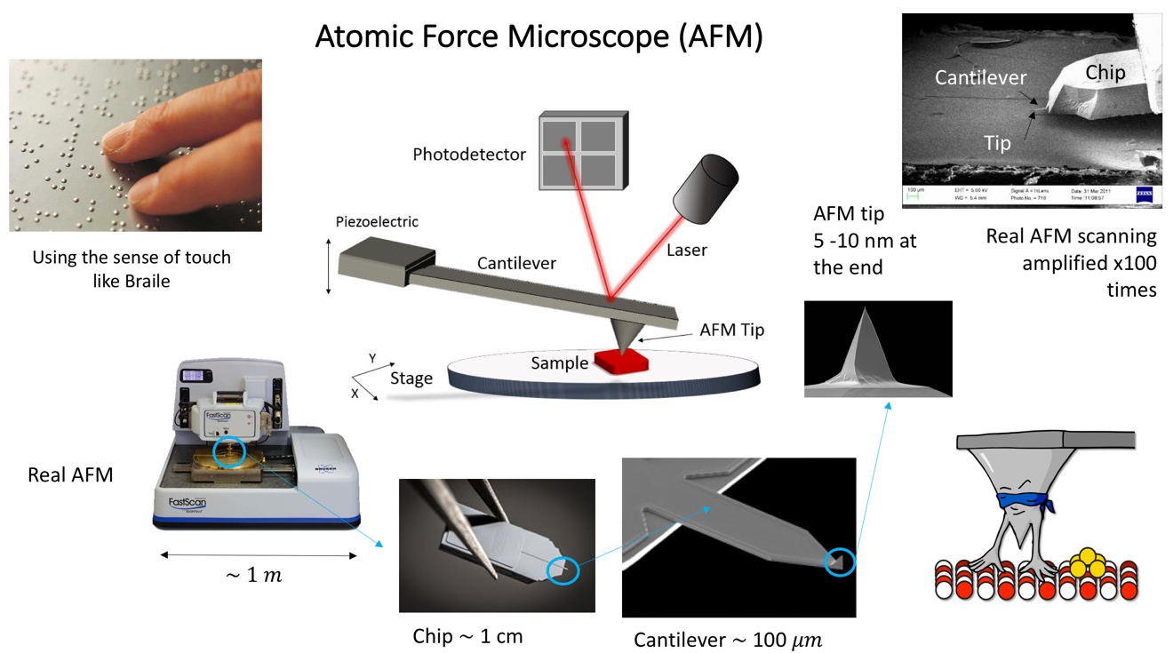
As humans, we ‘see’ because light interacting with the surfaces around us reaches our eyes. But how do we ‘see’ when want to study objects that are so small light steers around them? - like the study of objects on a nanometric scale such as individual proteins or molecules,  far below the resolution limit imposed by the properties of light?
​
One solution to this is to use an object to 'touch' or 'feel' a surface to detect  what shape it has. Basically, we ‘do Braille’!

Like Braille, the next logical sense to be used when the sense of sight is missing is the sense of touch. Unlike any other microscope, the Atomic Force Microscopy (AFM) was invented in 1980’s with this idea that if we have a ´finger´ small enough, we could be able to touch and study soft biological objects such as bacteria cells, tissues, proteins, nucleic acids, etc.

​The main advantage of this technique is that we use a nanometric tip to scan entire surfaces and obtain topography of the samples without transferring energy to the sample and hence performing no harm to biological samples. At the Florey Institute we collaborate among the Departments of Physics and Astornomy, MBB and Medical School at the Hobbs’ Lab to apply AFM to study DNA interactions with other proteins and the topography of Bacteria cells.   
​
Picture
​
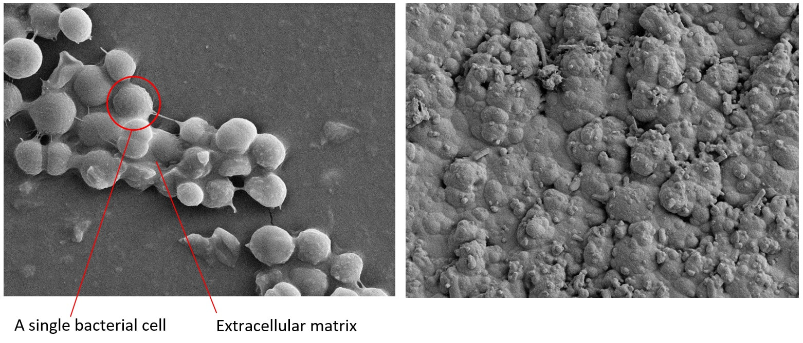
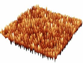
The landscape of the Bacteria Cell:
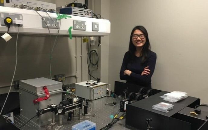
In the Hobbs’ group we are very interdisciplinary. That is why there has been a long-term collaboration between Prof. Jamie Hobbs from Physics and Prof. Simon Foster from Microbiology. Carrying out this project Laia Pasquina Lemonche, a third year PhD Cohort student is using the Atomic Force Microscope to study more in depth the topography of bacteria and the molecular organization of the cell wall from Staphylococcus aureus and Bacillus subtilis.

One of the main advantages of using this technique instead of other high resolution approaches is that we can obtain very detailed information from a single cell without the need for averaging any data. As a result, we can start answering fundamental questions about the morphology of bacteria and their behaviour but also new questions are arising from these findings. Through this, we are one step closer to better understand the host-pathogen interaction and how to treat bacterial infections effectively.
Picture
On the left - bacteria cell alive in 3D made by the AFM at 'low resolution'; On the right - 'high resolution' image of purified cell wall made by the AFM revealing the surface structures of the cell.

​DNA-protein interactions:
Another great application of AFM besides studying the topography of a sample, it is its ability to ‘video record’ biological reaction. For instance, DNA Polymerase, an enzyme responsible for DNA replication and repair during cell division, can be allowed to act on DNA, and consecutive AFM images of the process can be taken so as to ‘see’ the process actually happening in an environment mimicking the native cell conditions. In our lab, we are collaborating with Jon R. Sayers to use the AFM approach to understand the interaction between a DNA molecule and the Flap-endonuclease protein. Vinny Verma, a second year PhD Cohort student is using High-speed AFM to investigate this interaction to understand the conformation changes that occur during the process and how the cell manipulates the DNA using biomolecular machines.
​
For AFM the prospects are many, as we’re yet to see what new advancements  AFM can do for 'high resolution' imaging and even what video-AFM can do for our understanding of cells!
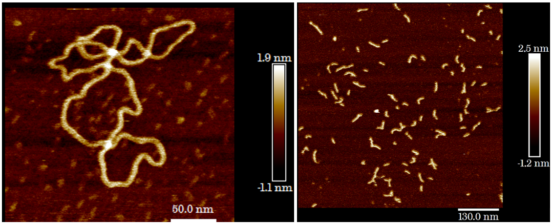
Picture
​Plasmid DNA imaged in air using AFM
Short DNA strands imaged in buffer using AFM
2 Comments

Day 9 - Seeing is believing: Developing chemical probes to image penicillin binding proteins

16/3/2019

0 Comments

 
​Haneesh Gangotra, a 3rd year PhD student with the Florey Institute, shares a brief overview on the multidisciplinary techniques he’s used for imaging penicillin binding proteins (PBPs).

Picture
​PBPs are essential enzymes used by bacteria to synthesise their cell wall and are the target of β-lactam antibiotics such as penicillin. Over time however, bacteria have formed new PBPs which resist these drugs and given rise to highly resistant strains such as MRSA.
​
​Being a chemist you would think I found the first part the easiest, that is synthesising suitable fluorescent probes which could bind to PBPs, but it was far from trivial! β-lactam’s are incredibly strained molecules and as such are difficult to manipulate. They can also be liable to degradation and this limits the type of chemistry you can apply. 
Picture
Structure of a penicillin derivatised chemical probe
​
​Luckily I eventually developed a protocol to work from and was able to successfully take these probes from the bench, apply to S. aureus and actually see them binding to PBPs using microscopy!
Picture
From synthesis and purification to application and imaging of PBPs in S. aureus: Development of a BODIPY based β-lactam chemical probe

​I have recently established a probe for using in Stochastic Optical Reconstruction Microscopy (STORM) which is something we’ve wanted for a while! This will allow crystal clear images of PBP interactions and offer an insight into their precise location during cell wall biosynthesis.
​
0 Comments

Day 8 - Early penicillin production - Dr Frank Ryan

15/3/2019

1 Comment

 
Picture
Frank Ryan is an emeritus consultant physician with an interest in medical and scientific history. He told us the story of a strange-looking vessel (pictures below) and its role in the early production of penicillin:

​"This ceramic vessel  was given to me by Norman Heatley, one of the pioneers of the penicillin story, when I interviewed him about his life in science in 1997. It was one of the vessels in which he produced penicillin at Oxford in the early trials before commercial production."

Picture
A ceramic vessel used for the early production of penicillin. Vessels were filled with media and inoculated with the penicillium mold which produced penicillin after eight days of growth.
Picture
For extraction and purification, air was blown into the vessels using a 'changing pistol' which forced air in and drew the culture medium, containing penicillin, out for further processing. ​(The full explanation of this process can be followed here: LINK)
Picture
Scaled production of penicillin using the culture vessels like those belonging to Frank Ryan. Image credit: http://www.penicillinstory.org
​
​"I first came into contact with Dr Heatley after I wrote a book, Tuberculosis: The Greatest Story Never Told, which detailed the history of the discovery of the cure for tuberculosis. In essence this was an integral part of the history of discovery of antibiotics and chemotherapy of infection. Dr Heatley made an important contribution to the initial production of penicillin in sufficient purity and quantity to allow the early trials of its production, safety, efficacy and potential to treat a range of different bacterial infections."

Picture
Dr Norman Heatley, one of the unsung pioneers involved in the the penicillin story.
Picture
Early designs for the culture vessels.

​"Heatley missed out on the Nobel Prize in Physiology or Medicine awarded in 1945 to Alexander Fleming, Howard Florey and Ernst Chain.  In the words of Sir Henry Harris, 'Without Fleming, no Chain or Florey: Without Florey, no Heatley; without Heatley, no penicillin.'"

​A fantastic video showing how early penicillin production was carried out and the role these ceramic vessels  can be found here: LINK
An archived video showing early production of penicillin can be found in the Wellcome Library: LINK
​
Ref: Harris H. Notes Rec. R. Soc. Lond., 1999; 53: 243-52
Image Credit: ​http://www.penicillinstory.org

 and https://blogs.bodleian.ox.ac.uk/archivesandmanuscripts/tag/penicillin/
1 Comment

Day 7 - Living without oxygen

14/3/2019

0 Comments

 
Natalia Hajdamowicz and Rebecca Hull work alongside Prof. Alison Condliffe investigating the impact of low-oxygen conditions on interactions between the human immune system and the infectious bacterium Staphylococcus aureus (also called MRSA).
Picture
Staphylococcus aureus has the propensity to develop anti-microbial resistance, causing a variety of infections which are becoming increasingly challenging to treat. In the Condliffe lab we use the SCI-tive Ruskinn hypoxic workstation (see photo below) to incubate cells in low-oxygen conditions equivalent to those encountered in our blood stream.

During infections S. aureus have evolved to circumvent our immune systems and this seems especially true in low-oxygen environments. Consequently, a deeper understanding of how our immune cells interface with the bacteria in real infection environments is essential.

In the Condliffe lab:

Natalia, a 3rd year Florey Institute PhD student, is looking at the mechanisms behind this host-pathogen interaction.

Rebecca, a 1st year MRF PhD student (
@MedResFdn), is looking at the evolutionary adaptations of these bacteria within this immune environment.

Exploring molecular mechanisms underlying host-pathogen interactions may lead to the development of successful treatment of intracellular infections by targeting host cell functions supporting bacterial growth or augmenting bactericidal functions.
Picture
Natalia (left) and Rebecca (right) growing cells in the SCI-tive Ruskinn hypoxic workstation, a low-oxygen cabinet that mimics oxygen conditions found in the human blood stream.
0 Comments

Day 6 - Developing structured antibacterial surfaces

13/3/2019

0 Comments

 
​As one of the interdisciplinary PhD students within the Florey cohort I get to split my time between the Physics and MBB departments. Coming from a physics background it can be easy to overlook the work that goes on in the other departments and so for me one of the best parts about my PhD is being able to hear about research and learn techniques from both disciplines.
​
Picture
​In my project I am aiming to develop antibacterial surfaces that are inspired by cicadas, who get their bacteria-killing ability from nano-scale pillars on the surface of their wings. The first part of my project is to study these wings with imaging techniques such as Atomic Force Microscopy and Scanning Electron Microscopy, to learn more about their nanoscopic features. I will then use this to design and make my own antibacterial surfaces and study how cells interact with them, in order to find out more about how these structures kill bacteria.​

​This collaboration between bacteriology and materials physics will hopefully provide a “drug-free” method of reducing the accumulation of bacteria on surfaces. This could eventually prove to be useful in a medical environment, especially in the fight against the growing issue of antimicrobial resistance.
​
Picture
0 Comments

Day 5 - Hero vs Villain: The Macrophage's Role in Cryptococcosis

12/3/2019

2 Comments

 
My name is Katherine Pline and I’m a third year PhD student in the Johnston Lab in Sheffield. I am interested in how invading pathogens and infections interact with the human immune system. I work on an infectious agent called Cryptococcus neoformans, a yeast which is inhaled into the lungs and can infect the brain.
​
Picture
5 day old zebrafish larva infected with C. neoformans (Kn99gfp  - green)

​When an infection occurs, human white blood cells are called to the scene to combat the infection. Like most events in life there is more than one way to go, and more than one possible outcome. If the immune system wins, the infection is defeated. If the immune system fails, the infection spreads. In C. neoformans infection, white blood cells called macrophages can either eat and kill the yeast, or can carry it to distant parts of the body and infect the brain. The direction the macrophage goes in depends on what happened to it before – its backstory.
​
Picture
The Johnston Lab
We have used zebrafish infections to watch how the infection progresses in each fish through time and, with help from the brilliant James Bradford and Dr Rhoda Hawkins, have used this to make a computational model of what happens in infection. This research has helped us to understand how the immune system interacts with the pathogen C. neoformans, how the yeast uses our macrophages to spread, and how our body mounts defences to stop it. Like the journeys we all face, infection here can lead to a happy ending, or can end in disaster and disease. While each person who faces a challenge, an infection, is unique, we must all fight to be victorious, and learn as much as we can in the process.
Picture
Human macrophages (MDMs) infected with C. neoformans  (Kn99gfp - green)
2 Comments

Day 4 - Testing new drugs to tackle corneal blindness

11/3/2019

6 Comments

 
Picture
As a first year PhD student, my research journey has only just begun. I’m based in the Department of Infection, Immunity & Cardiovascular Disease (IICD) and my focus is corneal infections.

The cornea is the transparent lens that covers the surface of our eyes and it plays an essential role in eyesight.  Therefore if bacteria infect the cornea, the results can be devastating and corneal infections are still a major cause of blindness worldwide. This type of infection is a particular problem in developing countries.

One of the major issues with treating corneal infections is that bacteria attach to the cornea as ‘biofilms’. A biofilm is a complex community of bacteria, held together in a sticky extracellular matrix. Bacteria within biofilms have different characteristics to free-living bacteria and one of these characteristics is an increased resistance to antibiotics. This makes it difficult to treat corneal infections and new antimicrobials are desperately required.

With this in mind, I am trying to develop an in vitro model of the infected cornea that can be used for drug testing. If successful, my PhD journey could end with the identification of new antimicrobials that are able to beat the biofilms and help to prevent corneal blindness.

Picture
An S. aureus biofilm:
These images were produced by my supervisor, Dr Rahaf Issa, using A Scanning Electron Microscope (SEM). The first image shows a biofilm early in its development and the second image shows a mature biofilm.


6 Comments

Day 3 - Corrigan Lab blog posts

10/3/2019

0 Comments

 
In the Corrigan lab, within the Florey Institute we focus on how bacteria, specifically methicillin resistant Staphylococcus aureus (also called MRSA), respond to and deal with stressful conditions such as starvation.

​​Bacteria like MRSA produce two molecules when stressed, collectively called (p)ppGpp, as part of the stringent response. This alters their physiology in many ways – including modulating the transcriptome, inhibiting the synthesis of ribosomes and generally slowing growth.
​

Picture
​The Corrigan Lab
​
​The lab as a whole is involved in studying many stages of the stringent response, ranging from the factors that trigger (p)ppGpp production and how the synthesis enzymes are regulated both transcriptionally and allosterically, to the cellular targets of (p)ppGpp and how they are affected, such as ribosome-associated GTPases and their interacting proteins.

​Recently, we have even branched out into the field of investigating potential antimicrobial surfaces based of the structure of cicada wings. To investigate these in more detail, we have been expanding into different specialisations and forging collaborations to increase our potential approaches– notably into biophysics, atomic force microscopy, enzyme kinetics, fluorescence studies and structural biology. Overall, the more detail we uncover regarding the stringent response, the wider our field of study seems to become.
​
Picture
Atomic Force Microscopy scan of the surface topology of a super-hydrophobic, antimicrobial cicada wing, showing the hexagonal array of nanopillars
Picture
Crystals of RsgA bound to ppGpp, which can be used to gain an X-ray diffraction pattern and solve the complexs structure to 0.19 nm resolution
0 Comments

Day 2 - Using advanced imaging techniques to understand host pathogen interactions

9/3/2019

2 Comments

 
Picture
My name is Yin Xin Ho and I am a 2nd year PhD student working under the supervision of Dr. Lynne Prince and Dr. Ashley Cadby. My project involves developing and using super-resolution microscopy techniques to understand neutrophil and Staphylococcus aureus interaction. Neutrophils are the most abundant white blood cells in our blood and are crucial immune defenders against ​S.aureus, a highly adaptive pathogen that causes wide range of infections. 

​​S.aureus have evolved ways to overcome neutrophil killing. One of these is to cause neutrophil to burst, thus escaping and causing infection elsewhere. The goal of my project is to understand how S.aureus disarms neutrophils in order to find ways to boost the host defence against this pathogen.

My project is highly interdisciplinary, which means that I spend my time between the Medical School and Biophysical Imaging Centre (BICEN) in the department of Physics and Astronomy. ​
Picture
A high-resolution image of neutrophil (red) containing S. aureus (green) taken using the Airyscan microscope in the Wolfson Light Microscopy Facility.

A typical day in medical school involves getting blood from volunteers in the morning to isolate neutrophils for my experiments. Over in BICEN, I work alongside physicists, engineers and chemists to build microscope, as well as to use the microscopes that are developed in the lab to image my samples.
​
Picture
​A density gradient to isolate different populations of blood cells
2 Comments

Day 1 - British Science Week 2019!

8/3/2019

0 Comments

 
Picture
Once again at the Florey Institute, we will be celebrating British Science Week with a series of blog posts over the next 10 days highlighting some of the exciting work that we do.

​​Inspired by the work of Sir Howard Florey, the main aim of the Florey Institute is to investigate infectious diseases and antibiotic resistance by looking at the interaction between pathogens and their hosts. This research is an interdisciplinary collaboration between scientists from across the University of Sheffield, with members from the MBB, APS, IICD, Chemistry and Physics departments as well as clinicians from the
​
Sheffield Teaching Hospitals. 


Keep revisiting this page each day over the course of the event to find out more about the research that our scientists do, as well as introductions to some of our research groups and other exciting news!

Follow the event on Twitter with #BSW19

Picture
0 Comments
    News Feed
    Upcoming Events
    Past Events
    ​
    Picture

    News + Events Archives

    April 2021
    March 2020
    January 2020
    December 2019
    August 2019
    July 2019
    June 2019
    May 2019
    April 2019
    March 2019
    January 2019
    June 2018
    March 2018
    January 2018

    2017 - 2015

Picture
Picture
The University of Sheffield 
Western Bank 
Sheffield, South Yorkshire 
S10 2TN 

United Kingdom
The University of Sheffield | Research | Collaborate | Team | Jobs | Cookies policy | Privacy
Powered by Create your own unique website with customizable templates.

BACK TO TOP

© 2015 The University of Sheffield

© DivTag Templates Ltd | All Rights Reserved