| Haneesh Gangotra, a 3rd year PhD student with the Florey Institute, shares a brief overview on the multidisciplinary techniques he’s used for imaging penicillin binding proteins (PBPs). |  |
PBPs are essential enzymes used by bacteria to synthesise their cell wall and are the target of β-lactam antibiotics such as penicillin. Over time however, bacteria have formed new PBPs which resist these drugs and given rise to highly resistant strains such as MRSA.
Being a chemist you would think I found the first part the easiest, that is synthesising suitable fluorescent probes which could bind to PBPs, but it was far from trivial! β-lactam’s are incredibly strained molecules and as such are difficult to manipulate. They can also be liable to degradation and this limits the type of chemistry you can apply.
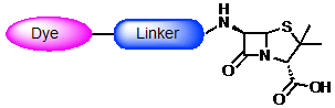
Structure of a penicillin derivatised chemical probe
Luckily I eventually developed a protocol to work from and was able to successfully take these probes from the bench, apply to S. aureus and actually see them binding to PBPs using microscopy!
Luckily I eventually developed a protocol to work from and was able to successfully take these probes from the bench, apply to S. aureus and actually see them binding to PBPs using microscopy!
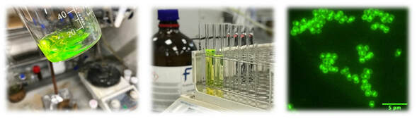
From synthesis and purification to application and imaging of PBPs in S. aureus: Development of a BODIPY based β-lactam chemical probe
I have recently established a probe for using in Stochastic Optical Reconstruction Microscopy (STORM) which is something we’ve wanted for a while! This will allow crystal clear images of PBP interactions and offer an insight into their precise location during cell wall biosynthesis.
