Current PhD students |

Kyle Buchan Email: [email protected] Department: Infection, Immunity & Cardiovascular Disease Supervisors: Prof. Steve Renshaw (IICD), Prof. Simon Foster (MBB) Project: Building an improved model for Staphylococcus aureus infection by humanising components of the Zebrafish immune system |
See also |
Staphylococcus aureus is one of the most important pathogens facing the world today. So far, much of our understanding of how S. aureus causes disease has been derived from experiments using infection models such as rabbits, rats and mice; however, these models can only offer so much without fully representing a typical human infection. Recently it has been found that many of the virulence factors produced by S. aureus are only active within humans, meaning that any infection model lacking the targets of these factors cannot account for their role during infection. The Zebrafish (Danio rerio) is already an established model in developmental biology and immunology research, with many tools and techniques readily available for working with them, facilitated by their genetic tractability. As well as being amenable to imaging and reproducible in high-throughput, Zebrafish have an immune system that is closely similar to humans, making it already a more suitable model for the study of infection over existing models. The project will aim to create transgenic lines of Zebrafish which express these human components that are specifically targeted by S. aureus; these lines can then be dissected with high resolution to determine some of the roles that these factors play in shaping a S. aureus infection. |
My project aims to use imaging techniques to describe the molecular and mechanistic events of phagocytosis. We hope to develop mulitplex imaging methods to analyse changes in the physical properties of a cell, as well as its cell membrane and actin components during early stages of phagocytosis. To acheive this we will use advances in superresolution microscopy, fluorescence labelling, and computational software. 
Emily Fisk Email: [email protected] Department: Infection, Immunity & Cardiovascular Disease Supervisors: Prof. David Dockrell, Dr Helen Marriott (IICD) Project: Priming macrophages for antimicrobial killing through pattern recognition receptor mediated metabolic changes |

Streptococcus pneumoniae remain one of the leading causes of infection-related death globally. Macrophages play a critical role in regulating pneumococcal replication in the airway and are essential in clearing bacteria from the lower respiratory tract. To mediate their antibacterial host defence roles macrophages need to be appropriately activated to optimize bacterial ingestion and killing. Pattern recognition receptors play a key role in optimizing bacterial macrophage activation and these shifts in phenotype are linked to shifting reliance on oxidative phosphorylation and glycolytic metabolism. The project will examine how different pattern recognition receptors influence metabolism and link this to generation of reactive oxygen species by mitochondria. The project will use a variety of approaches to analyse metabolic function and link these to features of macrophage polarization, generation of microbicidal molecules and antibacterial killing.
Penicillin and successive generations of β-lactam antibiotics have played a vital role in human healthcare for over 80 years; however their effectiveness has decreased markedly due to bacterial resistance. These antibiotics work by targeting penicillin binding proteins (PBPs) – a group of enzymes which are responsible for biosynthesising peptidoglycan, a unique and essential macromolecule present in bacterial cell walls. Bacteria have evolved over time creating new or modified PBPs which are less sensitive to β-lactam antibiotics. The exact mechanism of how this occurs is not understood, nor do we know how PBPs interact with their natural substrates. The project will aim to develop novel chemical probes to image PBPs, specifically PBP2a which is responsible for MRSA, using super resolution microscopy. This will help us understand: (i) the fundamental mechanisms of PBPs, (ii) the structural diversity of peptidoglycan, and ultimately (iii) an understanding of how β-lactam antibiotic resistance occurs.
Staphylococcus aureus (S. aureus) is a major cause of human death and disease but the factors that determine the outcome of this host-pathogen interaction are still poorly understood. Both bacterial colonisation and immune cell infiltration induce local tissue hypoxia; sites of infection are characterized by profound hypoxia, which may exert a dramatic effect on host-pathogen interactions. It has been shown that hypoxia impairs the ability of neutrophils to kill intracellular S. aureus but promotes the extracellular release of a range of anti-Staphylococcal proteins. Other immune cells such as Kupffer cells are key in determining whether S. aureus infection becomes disseminated, but the impact of hypoxia on these cell populations is unknown. We hypothesise that tissue hypoxia is a key determinant of the outcome of these host-pathogen interactions. The project will aim to explore this hypothesis, using primary human phagocytes derived from the peripheral blood of healthy volunteers or mouse liver. Hypoxia (3-12 KPa) will be delivered using a Ruskinn SciTive hypoxic workstation. |


Staphylococcus aureus is a highly successful pathogen that can circumvent many aspects of immunity. Neutrophils, the most abundant of our white blood cells, are a critical defence against infection and yet S. aureus is able to render them ineffective. S. aureus does this by rupturing an intracellular neutrophil compartment known as the phagolysosome, which is where bacteria are normally contained and killed. Escape from the inhospitable phagolysosome allows S. aureus to replicate inside the neutrophil cytosol, eventually bursting the neutrophil open and becoming free to cause infection elsewhere. This project will use super-resolution and confocal microscopy techniques to determine how S. aureus disables the immune response in this way, observing interactions of S. aureus with subcellular components of human neutrophils on a molecular level, visualising early sub-cellular trafficking of the bacteria, interactions with endogenous microbicidal factors, phagosome lysis and escape on a molecular level. The ultimate aim of this project is to identify ways of preventing escape of S. aureus from the phagosome, in order to improve the host defence against this multi-drug resistant and life-threatening microorganism. 
Sophie Irving
Email: [email protected] Department: Molecular Biology and Biotechnology (MBB) Supervisor: Dr Rebecca Corrigan (MBB) Project: Functional characterisation of (p)ppGpp synthases: essential enzymes required for bacterial stress adaptation and survival Staphylococcus aureus is a human pathogen responsible for a significant amount of disease and morbidity worldwide. When this bacterium invades a human host it encounters a number of different stresses, such as nutrient limitation. The bacteria respond to these stresses by switching on a response called the stringent response. This response results in the synthesis of two small nucleotides, collectively referred to as (p)ppGpp. These nucleotides are the effectors of the stringent response and function by binding to target proteins leading to the bacterial cells shutting down active growth and entering a slow growing or persistent state that promotes survival and leads to chronic infections. In S. aureus (p)ppGpp is synthesised by the bifunctional enzyme RSH and the monofunctional enzymes RelP and RelQ, however relatively little is known about the production and regulation of these synthases. This project will use molecular genetics to provide an in-depth characterisation of the environmental host signals that trigger production of these enzymes on both a transcriptional and translational level. Additionally this project will make use of an S. aureus mutant library to identify which regulator(s) control the transcription of these genes on a molecular level. Altogether this project will provide key insights into the synthesis of (p)ppGpp by these enzymes and so generate important mechanistic data on the pathogenesis of S. aureus.
The bacterial cell wall is essential for viability. The major stress-bearing cell wall polymer that can withstand the internal cellular turgor pressure in Staphylococcus aureus is peptidoglycan (PG). Because of this, PG synthesis is the target of antibiotics such as penicillin. 
Isuru Muthukudaarachchi
Email: [email protected] Department: Chemistry Supervisors: Dr Simon Whawell (Clinical Dentistry), Prof. Julia Weinstein (Chemistry) Project: Transition metal complexes as new antimicrobial-PDT agents Photodynamic (light-activated) therapy (PDT) is an emerging powerful approach to treat illnesses. It uses special drugs which are only toxic when illuminated by visible light, but remain non-toxic without light. This approach presents tremendous opportunities for targeted therapy and dramatically reduce side-effects. So far, the main focus of research in this area was on killing cells – especially cancer cells. This project aims at an even more ambitious target – we want to translate our recent developments in anti-cancer PDT to kill bacteria. Why is it such a challenge? Because bacteria invade the cells, and one must kill bacteria, but not the host cell.
|


Streptococcus pyogenes or Group A Streptococcus (GAS) is capable of causing severe and lethal diseases such as necrotising fasciitis and Streptococcal Toxic-Shock Syndrome and globally accounts for over half a million deaths a year. Remarkably in this day and age of antimicrobial resistance GAS remains sensitive to penicillins and cephalosporins yet recurrent infections and treatment failures are common. One theory as to why this recurrence occurs is due to the formation of biofilms by GAS which may help to shield against antimicrobials and aid in host immune evasion. A common component of many bacterial biofilms are functional amyloid proteins, which utilise the inherent biophysical properties of amyloids to reinforce and provide robustness to the biofilm matrix.
Atomic force microscopy (AFM) is a powerful tool that allows us to measure local mechanical properties, enabling us to image living systems at the nanometric scale. I aim to use and develop these capabilities to study how bacteria (e.g. Staphylococcus aureus) make their cell walls, and how this process is inhibited by β-lactam antibiotics (e.g. penicillin). With antimicrobial resistance (AMR) on the rise, research into how antibiotics kill bacteria is essential for the discovery of novel and promising solutions. This project will be carried out in Jamie Hobbs' lab in collaboration with Simon Foster's group. |

Streptococcus pyogenes, also known as Group A Streptococcus (GAS) is capable of causing a seemingly boundless diversity of infections, ranging from the relatively self-limiting; impetigo, scarlet fever and pharyngitis, to the more severe and potentially lethal necrotising fasciitis and toxic shock syndrome. GAS is also responsible for considerable morbidity and mortality due to post-infectious immunological sequelae, predominantly rheumatic heart disease and glomerulonephritis. 
Daria Shamarina Email: [email protected] Department: Molecular Biology and Biotechnology (MBB) Supervisors: Simon Foster (MBB) Project: Interactions between S. aureus components and the host immune system |

Staphylococcus aureus is a wide-spread pathogen that can be acquired from the community or in hospitals. Though serious infection is uncommon in healthy and young individuals, it can cause septic arthritis, pneumonia and endocarditis amongst other diseases and carries a high mortality rate despite treatment. Importantly, S. aureus can also be resistant to many antibiotics. Therefore, increased understanding of the interaction of the pathogen and its host may lead to new control regimes. Previous research has shown that a combination of various bacterial components are able to interact with the host immune system and cause septic shock leading to death. The aim of this project is to determine the S.aureus components that are responsible for causing immune activation in the host. In the long-term this will benefit the development of novel anti-infection strategies.
Flap endonucleases (FENs) play a vital role in DNA replication, repair and recombination in all living cells. Aspects of how FENs locate their branched DNA substrates and the conformational changes associated with DNA hydrolysis remain unclear. In my project, we are trying to address these questions using a combination of direct atomic force microscopy (AFM) imaging with conventional biochemical and structural approaches. Development of imaging of DNA to major and minor groove resolution using approaches initially proven for membrane protein imaging, has provided the potential to directly visualize local strain within a single molecule during an interaction with a bound protein. Using AFM we aim to explore how FENs engage their substrate using imaging of DNA-FEN complexes to characterize their hydrolysis.
Streptococcus pneumoniae is a significant global pathogen and a leading cause of community acquired pneumonia. In order to cause infection, S. pneumoniae must overcome the plethora of defence mechanisms the innate immune system possesses, including antimicrobial action by lysozyme. |


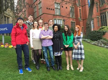
The first two cohorts of Florey students
|
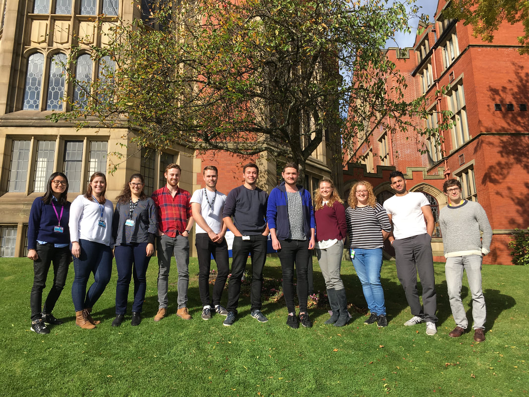
The current Florey students
|